Publications
Below you can find a selection of highlighted publications of our group. A full publication list can be found here:
https://www.rug.nl/staff/r.schirhagl/research/publications.html
Recent review articles about diamond based quantum sensing can be found here:
Nanoscale MRI for Selective Labeling and Localized Free Radical Measurements in the Acrosomes of Single Sperm Cells

Free radicals play a key role in sperm maturation. Here we were able to localize where this radical formation is coming from with nanoscale resolution. We have achieved that by measuring radical formation with nanodiamonds attached to the acrosome area for boar sperm cells.
Diamond Quantum Sensing Revealing the Relation between Free Radicals and Huntington's Disease

Huntington disease is a conditions where protein aggregation as well as free radical generation play a role. Here we investigate the free radical generation with sub-cellular resolution. We found that there is indeed free radical generation at the sites where protein aggregation occurs as well.
Quantum Sensing of Free Radicals in Primary Human Dendritic Cells

For unknown reasons certain people have more aggressive dendritic cells than others. Here we investigated if these donors also have a different free radical response. These are also the first relaxometry measurements that have been done on primary cells.
Applying NV center-based quantum sensing to study intracellular free radical response upon viral infections

Free radical generation plays a role in viral infections as well as in how our immune systems defends us against them. By conjugating nanodiamond sensors to viruses we were able to show the local radical load at the site of virus entry during infection with the semliki forest virus (SFV).
Quantum monitoring of cellular metabolic activities in single mitochondria

Here we performed free radical detection using diamond magnetometry measurements in single organelles. We were able to measure both in isolated mitochondria as well as mitochondria within cells. We see clear differences when we target nanodiamonds to mitochondria than when they are free in the cytosol when we trigger or inhibit free radical production in mitochondria.
The Shape and crystallographic orientation of nanodiamonds

While many researchers have assumed a spherical (or somewhat more isotropic) shape for fluorescent nanodiamonds, we found that they are actually more flake like. In addition, we could show that they have a preferred crystal orientation. This finding has important consequences on the sensing performance as well as chemical and biological properties.
How to get nanodiamond into cells?

While essentially no cytotoxicity for mammalian cells has been reported for nanodiamonds we did find surprising effects on bacteria. However, the effect is not that simple. The effect strongly depends on the medium as well as on the bacteria type. While we see 90% reduction of colony count for one type of medium we see reduction at all in a different medium. This study suggests that one needs to be very careful when using this for comparing nanoparticle toxicities. Later we found that this is due to bacterial aggregation rather than toxicity.
Aggregation of Nanodiamonds in cell media

We found that nanodiamonds aggregate severely in presence of cellular media. We identified proteins and salts, which are involved in this process. Finally, we also suggest a very simple process of reversing the order of mixing medium components with diamonds to avoid this problem.
Nanodiamond Relaxometry-Based Detection of Free-Radicals from Chemical Reactions in Biologically Relevant Conditions
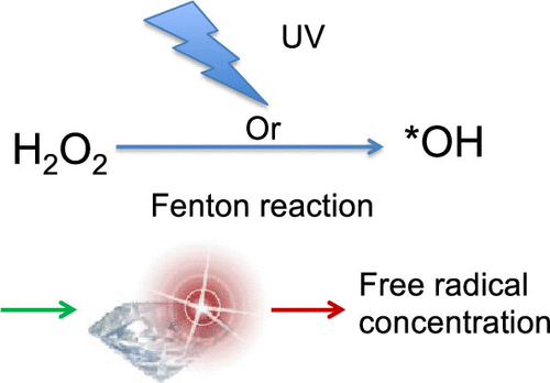
Relaxometry is a powerful tool for determining the free radical concentration in biological samples. Here we optimise the protocol and provide calibration measurements which allow us to translate a T1 response into a radical concentration.